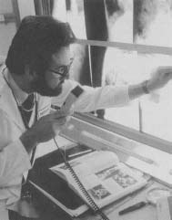
X-ray
X-rays are electromagnetic waves, like light waves, but with a wavelength about 1,000 times smaller. Because of this very short wavelength, X-rays can easily penetrate low-density material, such as flesh. They are reflected or absorbed, however, by high-density material such as bone. The picture made by an X-ray machine shows the denser materials (like bones) as dark areas.
X-Ray Discovery
In 1895 German physicist Wilhelm Roentgen (1845-1923) was experimenting with a cathode ray tube. The tube produced weak rays that caused a screen to fluoresce (glow). To create a controlled environment, Rontgen placed the cathode tube in a black cardboard box that was too thick for cathode rays to penetrate. Once the cathode ray tube was turned on, however, he noticed that another screen across the room began to glow. Since this second screen was too far from the tube for cathode rays to reach, especially through a layer of cardboard, Roentgen realized that he had discovered a new type of ray.
Through experimentation Roentgen found that this new ray was able to penetrate even the thick walls of his laboratory. Roentgen delivered a paper detailing his findings on December 28,1895. In the paper he admitted that he did not know the precise nature of these new rays. He chose to name them "X-rays," since "X" is the mathematical symbol for the unknown.
Few discoveries have been accompanied by as much fanfare as the X-ray. During the 12 months following the publication of Roentgen's paper, more than 1,000 books and articles were written on the subject. The number of publications rose to more than 10,000 before 1910.
A Diagnostic Tool
The penetrating power of X-rays to reveal bone structure was immediately recognized as a new medical diagnostic tool. Not all the excitement was positive, however. Many people considered the X-ray machine's ability to look through walls and doors an end to privacy. In fact, opera houses banned the use of X-ray binoculars in order to prevent patrons from peering beneath the actresses' costumes. Nevertheless, more rational minds eventually prevailed. Roentgen was awarded the first Nobel Prize for Physics in 1901.
Practical Uses of X-rays
The first medical use of X-rays came in 1896. It was American physiologist Walter Bradford Cannon who used a fluorescent screen to follow the path of barium sulfate through an animal's digestive system. This was possible only after Thomas Alva Edison invented the X-ray fluoroscope that same year. Soon after, physicians worldwide began using X-rays on humans, usually to examine bone fractures or to search for foreign objects such as bullets.

By 1970 most Americans were receiving at least one X-ray exam every year from physicians and dentists. However, recent evidence has shown that overexposure to X-rays can lead to the development of leukemia. Many doctors now recommend X-ray exams only when absolutely necessary
Ironically, the harmful side effects of X-ray scanning have suggested yet another use for the procedure called radiotherapy. In this therapy, very high frequency X-rays ("hard rays") are used to destroy cancer cells. Radiotherapy is most often used in conjunction with chemotherapy (cancer medicine that is taken by mouth).
X-Rays in Everyday Life
One of the most familiar X-ray machines is the baggage scanner found at airport terminals. This low-power X-ray device is placed over a conveyor belt, where it scans passengers' luggage. The machine used in this type of scanner must operate at a very specific frequency. It must be high enough to penetrate hard-shell baggage but low enough to prevent the accidental exposure of camera film.
[See also Barium ; X-ray crystallography ; X-ray machine ]
Thank you!
Thanks again:D