X-ray machine
The first X-ray device was discovered accidentally by the German scientist Wilhelm Roentgen (1845-1923) in 1895. He found that a cathode-ray tube emitted invisible rays that could penetrate paper and wood. The rays caused a screen of fluorescent material several yards away to glow. Roentgen used his device to examine the bone structure of the human hand. This
Upon their discovery in 1895, X-rays were advertised as the new scientific wonder and were seized upon by entertainers. Circus patrons could view their own skeletons and were given pictures of their own bony hands wearing silhouetted jewelry. Many people were fascinated by this discovery. Some people, however, feared that it would allow strangers to look through walls and doors and eliminate privacy.
Medical Use of X-rays
The most important application of the X-ray has been its use in medicine. This importance was recognized almost immediately after Roentgen's findings were published in 1895. Within weeks of its first demonstration, an X-ray machine was used in America to diagnose bone fractures.
What Are X-rays?
X-rays are waves of electromagnetic energy. They behave in much the same way as light rays, but at much shorter wavelengths. When directed at a target, X-rays can often pass through the substance uninterrupted, especially when it is of low density. Higher density targets (like the human body) will reflect or absorb the X-rays. They do this because there is less space between the atoms for the short waves to pass through. Thus, an X-ray image shows dark areas where the rays traveled completely through the target (such as with flesh). It shows light areas where the rays were blocked by dense material (such as bone).
Thomas Alva Edison invented an X-ray fluoroscope in 1896. American physiologist Walter Cannon used Edison's device to observe the movement of barium sulfate through the digestive system of animals and, eventually, humans. (Barium sulfate is a fine white powder that is still used as a contrast medium in X-ray photography of the digestive tract.) In 1913 the first X-ray tube designed specifically for medical purposes was developed by American chemist William Coolidge (1873-1975). X-rays have since become the most reliable method for diagnosing internal problems.
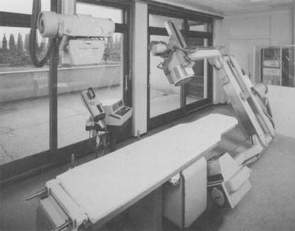
Modern X-Ray Machines
Modern medical X-ray machines have been grouped into two categories: those that generate "hard" X-rays and those that generate "soft" X-rays. Soft X-rays are the kind used to photograph bones and internal organs. They operate at a relatively low frequency and, unless they are repeated too often, cause little damage to tissues.
Hard X-rays are very high frequency rays. They are designed to destroy the molecules within specific cells, thus destroying tissue. Hard X-rays are used in radiotherapy, a treatment for cancer. The high voltage necessary to generate hard X-rays is usually produced using cyclotrons or synchrotrons. These machines are variations of particle accelerators (atom smashers).
One of the more familiar X-ray machines is the security scanner used to examine baggage at airports. These machines use a very low-power scanner. They illuminate the interior of purses and suitcases without causing damage to the contents.
[See also Bragg, William Henry and William Lawrence ; Insulin ; Penicillin ]
I would like to know how to take a chest X-ray to a pregnant woman. Should the lead shield place at the back or in the front of her body?
The reason i am aksing is one of my friend was asked to take X-ray while pregnant, and the lead shield was place at the back? I wonder the if X-ray beam emmitted from the back?
Rgds,
Gordon
thanks again
Thanks
this is amazing!
thanks a lot for the great help
I have an exam and this is perfect study material
thanks a million
From Sachi Blossoms Of Quezon National High School
you for those who made it!
This website is from another World.
From
Warri,Delta State, Nigeria.
Thanks
Thanks
"Dear Doctor,
I would like to know how to take a chest X-ray to a pregnant woman. Should the lead shield place at the back or in the front of her body?
The reason i am aksing is one of my friend was asked to take X-ray while pregnant, and the lead shield was place at the back? I wonder the if X-ray beam emmitted from the back?
Rgds,
Gordon"
*To put it in the simplest form possible without using specific radiology terms: There are two parts of an xray machine-- the tube (where the light comes from) and the wall bucky (box on the wall). The xrays come from the xray tube (light source) ; therefore, if the front of her chest was against the wall bucky, then the xray apron should've been on the back of her and if she was facing the xray tube (light on the front of her chest) then the apron should've been on her stomach. In short, the apron should be on the side of the xray tube (light source) because this is where the radiation comes out. :)