Prenatal diagnostic techniques
Prenatal diagnosis is the process of determining the condition of a fetus (name given to unborn young from the end of the eighth week of development through birth) before it is born. This type of diagnosis has become an important part of pregnancy care.
The earliest form of prenatal testing was very simple. The mother noted fetal activity in the womb, and the doctor manually felt the unborn child through the mother's abdomen. Eventually machines were developed for listening to the fetal heartbeat.
Amniocentesis
Prenatal diagnosis began to improve with the development of amnio-centesis in the 1950s. In the 1960s the ability to culture cells from amniotic fluid was developed. Beginning in 1968, cells from amniotic fluid could be analyzed for chromosome disorders like Down's syndrome. Today, amniocentesis permits the diagnosis of a wide range of disorders. Amniocentesis is normally recommended at fourteen to sixteen weeks of pregnancy. Test results are usually available to the patient in two to three weeks.
Ultrasound
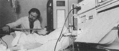
Ultrasound scanning has become the most popular method of prenatal testing. Ultrasound uses sound waves to produce a picture of the developing fetus. It has no known harmful effect on the mother or the unborn child and requires no probing or biological samples to be taken.
In 1977 so-called "real-time" ultrasound equipment became commercially available. The real-time mode shows detailed images of the moving fetus as the movement occurs. Real-time ultrasound was quickly applied to invasive (entering the body) prenatal diagnostic techniques. With
Chorionic Villus Sampling (CVS)
Chorionic villus sampling is when samples of fetal tissue are retrieved from the fetus. The samples are then analyzed for chromosomal, biochemical, and DNA content. One method of obtaining the sample through the cervix was described by Jan Mohr (1921-) in 1968. This method was abandoned because of a high rate of complications.
In 1982 a British group at the University College Hospital of London developed a new technique. Transcervical CVS sampling (taking a sample through the cervix) guided by real-time ultrasound became a safe procedure. Transabdominal CVS sampling (taking a sample through the abdomen) was first reported in 1984 and is used in some cases to avoid vaginal infection. Ultrasound-guided CVS is now widely used. It can be performed much earlier in pregnancy than amniocentesis and results are available within a week.
Measuring Alpha-Fetoprotein (AFP)
Another prenatal diagnostic test now given to most pregnant women measures maternal serum alpha-fetoprotein (MSAFP) levels. This test samples the mother's blood for the amount of AFP. AFP is a protein produced by the fetal liver. As levels of AFP in the mother's blood mirror the levels in the fetus, testing has been rapidly expanded to include more women. In 1984 it was reported that low MSAFP levels were associated with Down's syndrome. With this information, the need for more detailed testing can be identified earlier during the pregnancy.
Fetal blood sampling has been done since 1972. The sample is used to diagnose a variety of hereditary blood disorders. This procedure was improved upon by Fernand Daffos and his team in 1982. The group developed a method of retrieving samples of blood from the umbilical cord. They used a needle guided by real-time ultrasound imaging. This procedure is called cordocentesis, or percutaneous umbilical blood sampling (PUBS).
DNA Analysis
DNA analysis for specific gene disorders was introduced to prenatal testing in 1976. DNA from a number of different sources is examined. These sources include fetal samples plus maternal and paternal blood samples. This allows the doctors to identify genetic problems that might be transmitted by either parent. Over 260 single gene defects can now be diagnosed through DNA analysis, and the list continually grows.
Other New Procedures
Prenatal diagnostic testing is rapidly expanding. Nuclear magnetic resonance imaging ( MRI ) and spectroscopy (NMR) are new tools. The tests reveal biochemical information about fetal soft-tissue and organ structure and function. Fetal tissue sampling is an experimental technique. It is used to detect very rare hereditary and fatal skin disorders. Another not-yet-perfected experimental procedure is cell sorting. In cell sorting, fetal cells are isolated from the maternal blood for analysis. Prenatal testing is even carried out on some embryos, eggs, and sperm before in-vitro implantation.
After Diagnosis
Prenatal diagnosis is now moving in the direction of prenatal treatment. The field of prenatal surgery is in its infancy. A few conditions identified prenatally can be treated in the womb successfully by medicating the mother. Respiratory distress syndrome is one of these conditions. It is a prime killer of prematurely born infants. Another condition is methylamalonic acidemia. This is a rare vitamin metabolism defect.
I want to know if a fetus genotype can be check in 7 to 8 weeks of a pregnancy and where it can be checked in Nigeria
Thank you as I await your response.