Computerized Axial Tomography
A computerized axial tomography (CAT) scan is a special type of X-ray used for viewing the internal organs of patients. Although a regular chest X-ray can show the heart and lungs, the CAT scan can show the same organs but with detail 100 times greater and with little or no additional irradiation (exposure to radiation).
In the late 1960s Alan Cormack, an American physicist, and Godfrey Hounsfield, an English electrical engineer, independently developed a way to produce a three-dimensional image of the parts of the body, (called tomogram) by taking many different X-ray cross sections and combining them. Eventually, the two would share a 1979 Nobel Prize for medicine for their work, research which led to development of the CAT scanner.
Cormack was the first to construct a machine to "shoot" tomograms. His first model used a thin beam of X-rays aimed at one part of the body but taken from many different angles. Unfortunately he lacked a system that could process all of the information that one CAT scan produces. The solution to this problem was the computer. Hounsfield began working on his own CAT scanner in 1967. He used a system similar to Cormack's, but he also used a computer. The computer was able to sort all of the X-ray data into a picture. The first CAT scanner took nine days to complete a picture of a preserved human brain. Before it could be used for patients, Hounsfield had to improve its design. Later models required nine hours and eventually only nine minutes. His final apparatus took about five minutes to complete its scan. The first CAT machine of practical use was installed at Atkinson Morley's Hospital in Nimbledon, England, in 1971.
Today the CAT scanner finishes a scan in a few minutes. The pictures can be seen almost immediately on a television monitor, and within 30 minutes the entire scan can be copied to a computer disk. The scan can be
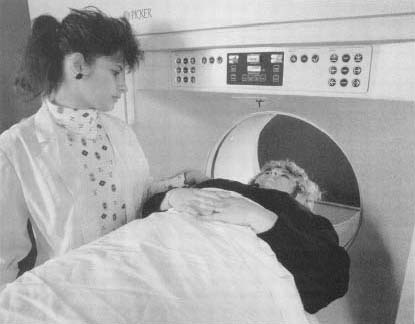
Scanning Procedures
The CAT scanner consists of a flat table on which a patient lies. A tube-shaped device is on either side of the table and is fitted with many X-ray scanners. Before the scan begins, the part of the patient that is to be scanned is moved inside this tube. The CAT scanner rotates 180 degrees around the patient's body and sends out a very thin beam of X-rays. Each X-ray scanner has a detector placed exactly opposite to it. The scanner and detector are built to move together so that when the scanner moves, it is aimed directly at the detector.
During the CAT scan, the scanner sends out a short impulse that is picked up by the detector. After the detector picks up the first signal, the scanner and detector rotate slightly and the same picture is taken again but at a slightly different angle. As the beam travels from the scanner through the patient, it is changed as it goes through bones and organs. The beam that the detector picks up is different from that the scanner emitted. This difference is noted by the computer. It analyzes this data and converts it into electrical impulses that make a picture on a television screen. The amount of data is huge, since some CAT scanners use up to 300 X-ray scanners taking 300 pictures each. This results in almost 90,000 X-ray slices or tomograms that the computer must convert into a picture.
In the 1970s a more advanced tomography technique was developed by Michael Phelps and Edward Hoffman, biophysicists at the UCLA School of Medicine. The duo developed a PET scan (PET is an acronym for positron emission tomography). For this scan, the patient has a radioactive material injected into the bloodstream. This radioactive material is not harmful and it emits positrons, a kind of beta particle (high-speed electron), and gamma rays. The PET scanner reads gamma rays like the CAT scanner reads X-rays. Since certain radioactive material travels to certain parts of the body (for example, iodine goes to the thyroid), more detailed pictures of a specific organ can be made.
Scanner Enhancements
CAT scanners have revolutionized the way doctors diagnose and treat patients. Since doctors can now see inside the patients body, the patient is often spared surgery. If the patient has a cancer in a certain part of the body and surgery is needed, the surgeon will have a clear idea of how big the cancer is, the surrounding tissues it may be affecting, and how much to cut out. If a cancer cannot be cut out, but it can be treated with X-rays, the CAT scanner can precisely map out the exact borders of the cancer and direct the X-rays to only the cancer.
CAT scanners are being continually refined to take pictures faster and faster. This is important for taking pictures of moving things like blood in a vein or artery. Scanners can now detect blockages, called clots or thromboses, in the arteries or veins. Most recently, an ultrafast type of CAT scanning has been developed called EBCT, an acronym for electron beam computed tomography. With this technique a scan can now be taken of the arteries of the heart, called coronary arteries. The scan reveals a cross section of the coronary arteries and shows the amount of calcium in the walls of the arteries. If there is too much calcium in the artery walls, it can cause a blockage that will lead to a heart attack. If these blockages are detected early, a heart attack can be prevented by doing a simple operation.
[See also X-ray machine ]
Comment about this article, ask questions, or add new information about this topic: